Get Premium
Dark mode theme is available exclusively for premium users. Learn more about the benefits of subscribing.
No fees, cancel anytime.

 Dark Mode Ad-Free Browsing Unlimited Content
 Ad-Free Browsing Unlimited Content Dark Mode
Join 1.2 million Panda readers who get the best art, memes, and fun stories every week!
Nikon has just announced the winners of its annual Small World Photomicrography competition, and as you can see from these stunning photographs, bigger isn't always better.
The competition is in its 42nd year and this year over 2000 people from 70 countries entered. For those that don't know, photomicrography is the practise of taking a photograph through a microscope or similar magnifying device in order to capture the intricate details of things invisible to the human eye. From the proboscis of a butterfly and the foot of a beetle to espresso coffee crystals, the pictures below give us a whole new way of looking at world. The categories are divided into winners, honorable mentions, and images of distinction, and you can find the full list on the Nikon Small World website.
More info: Nikon Small World (h/t: demilked)
This post may include affiliate links.
A few hundred years ago, biologists had to actually draw what they saw beyond the naked eye. Whether its 10X or 300X, the view under microscopes is mesmerizing.
A few hundred years ago, biologists had to actually draw what they saw beyond the naked eye. Whether its 10X or 300X, the view under microscopes is mesmerizing.


 No fees, cancel anytime


 Of A Male Diving Beetle")


 Crystals")


 Carefully Arranged By Hand In Victorian Style")


 Seed")


")


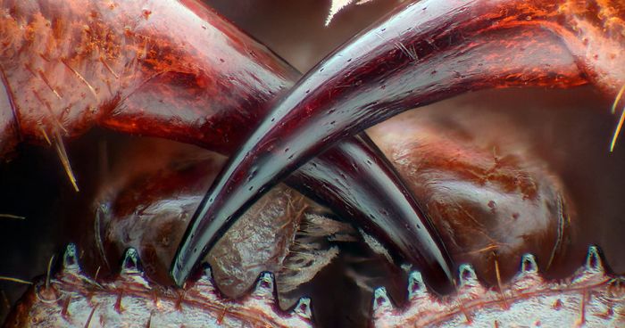
 Of A Tiger Beetle")